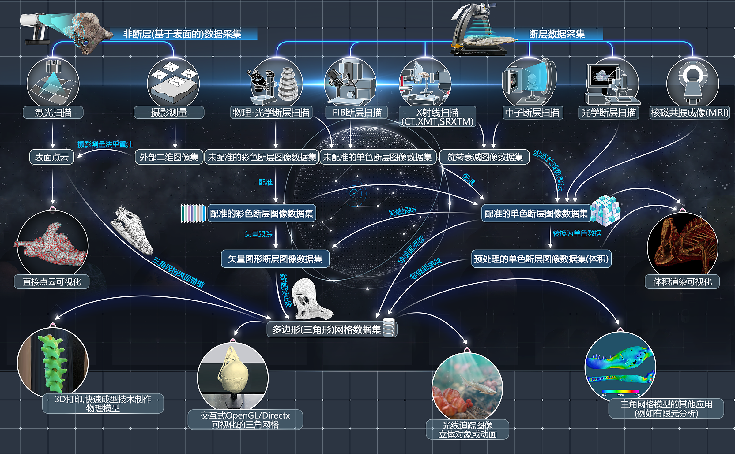


Archives of Digital Morph is a project-based data archive that allows researchers to store and organize, share, and distribute their own 3d data. Furthermore any registered user can immediately search for and download 3d morphological data sets that have been made accessible through the consent of data authors.
The goal of Archives of Digital Morph is to provide rapid access to as many researchers as possible, large numbers of raw microCt data and surface meshes representing vouchered specimens.
File formats include stl, tiff, raw, and ply. The website is designed to be self explanatory and to assist you through the process of uploading media and associating it with meta data. If you are interested in using the site for your own data but have questions about security or anything else contact the site administrator. Otherwise please download whatever data you need and check back frequently to see what's new.

160KV Micro-Computed Laminography system
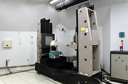
450 kV industry-computerized tomography
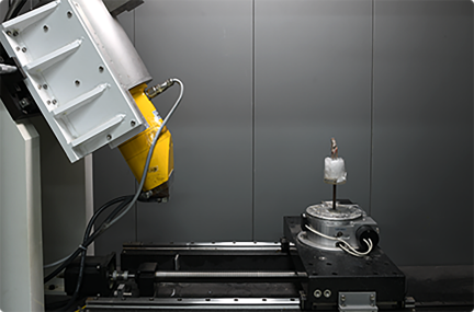
225 kV micro-computerized tomography
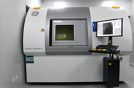
GE v|tome|x m300&180 micro-computed-tomography scanner
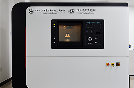
SNCT-800 Specral Micro-CT
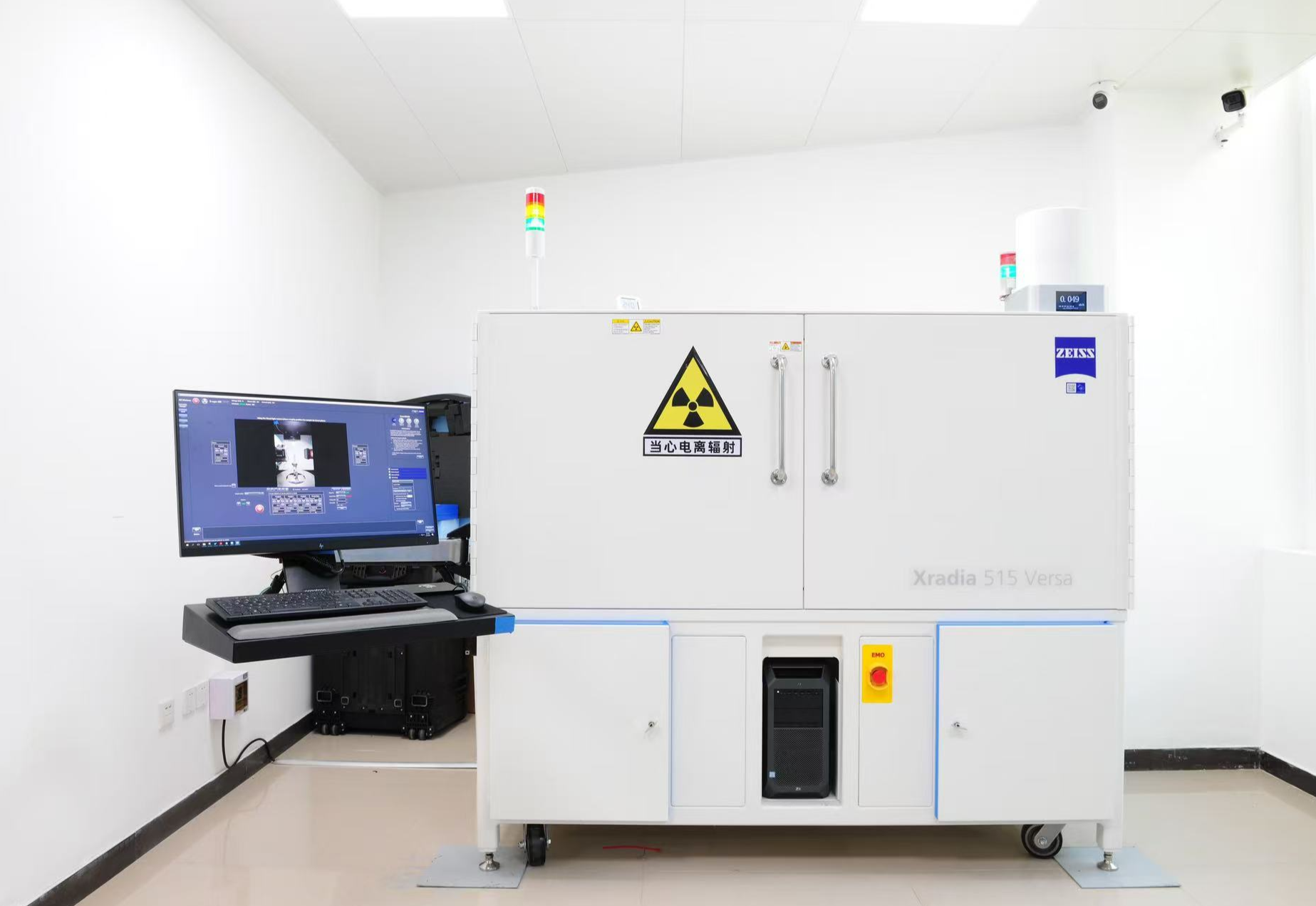
Xradia 515 Versa